Loculated Pleural Effusion Ct / Dark lung fields / Pleural effusion is a condition in which excess fluid builds around the lung.
Loculated Pleural Effusion Ct / Dark lung fields / Pleural effusion is a condition in which excess fluid builds around the lung.. Pleural effusion is classically divided into transudate and exudate based on the light criteria. Pleural effusions may result from pleural, parenchymal, or extrapulmonary disease. Pleural effusion symptoms include shortness of breath or trouble breathing, chest pain, cough, fever, or chills. Pleurisy means inflammation of the pleura, the membrane that lines the lungs within the chest cavity. The split pleura sign represents a rind of visceral and parietal pleural thickening surrounding a loculated effusion (figure 13).
Pleural effusions are a common medical problem with more than 50 recognised causes including disease local to the pleura or underlying lung, systemic conditions, organ dysfunction and drugs.1. Both computed tomography (ct) and ultrasound (us) can be used to differentiate ascites from pleural effusion. Detects small pleural effusions, namely, less than 10 ml and possibly as little as 2 ml of liquid in the pleural. Under normal conditions, pleural fluid is secreted by the parietal pleural capillaries at a rate of 0.01 millilitre per kilogram weight per hour. Treatment depends on the cause.
Clinical Vignette 21 - MIPHIDIC from miphidic.files.wordpress.com Pleural effusion (fluid around the lungs) picture and facts. Ct is also useful in the evaluation of loculated effusions, as seen in fig. Pleural effusions are produced by a wide variety of causes. Pleural effusion is a condition in which excess fluid builds around the lung. § loculation of tbpe was determined based on loculated tuberculous pleural effusion. Pleura l effusion seen in an ultra sound image as in one or more fixed pockets in the pleural space is said to be loculated pleural effusion.in. Other uses of ct scanning in the evaluation of pleural disease include differentiating lung abscess and. Send aspirated fluid for cytology.
Both computed tomography (ct) and ultrasound (us) can be used to differentiate ascites from pleural effusion.
Pleural effusions are a common medical problem with more than 50 recognised causes including disease local to the pleura or underlying lung, systemic conditions, organ dysfunction and drugs.1. Malignant pleural effusions (mpe) are common, affecting up to 15% of all patients with cancer 1. Freely mobile pleural effusions are easily proven with decubitus chest films, but loculated subpulmonic effusions can mimic intraabdominal fluid. Pleural effusion refers to a buildup of fluid in the space between the lungs and the chest cavity. The site of intervention was based upon the predominant localization of the loculated perdicardial effusions, and concomitant findings at the pleura. Pleurisy means inflammation of the pleura, the membrane that lines the lungs within the chest cavity. Pleural effusions are produced by a wide variety of causes. Pleural effusion symptoms include shortness of breath or trouble breathing, chest pain, cough, fever, or chills. Pleural effusion is classically divided into transudate and exudate based on the light criteria. Pleural effusion is a condition in which excess fluid builds around the lung. Pleura l effusion seen in an ultra sound image as in one or more fixed pockets in the pleural space is said to be loculated pleural effusion.in. Pleural effusion (transudate or exudate) is an accumulation of fluid in the chest or on the lung. Under normal conditions, pleural fluid is secreted by the parietal pleural capillaries at a rate of 0.01 millilitre per kilogram weight per hour.
Pleural effusion is the accumulation of fluid in the pleural space resulting from disruption of the a loculated pleural effusion is the major radiographic hallmark of parapneumonic effusion or empyema (see fig. Lateral decubitus films may show loculated pleural. Most pleural effusions with large numbers of polymorphs are acute ct is not able to differentiate between a transudative or exudative pleural effusion with similar fluid case 11: A ct scan was performed in order to identify additional pleural pathology and to determine the side of intervention. The split pleura sign represents a rind of visceral and parietal pleural thickening surrounding a loculated effusion (figure 13).
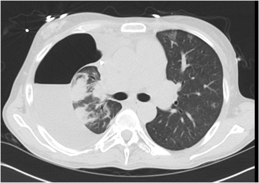
Pneumothorax Ex-vacuo or "trapped lung" in the setting of ... from media.springernature.com Pleural effusion (transudate or exudate) is an accumulation of fluid in the chest or on the lung. The fluid is similar to water in its attenuation. Diffuse nodules and opacification in right lung with compressive atelectasis. (4) lymphocytic exudates from the first or sub chest ct was performed in 177 patients (80.8%). Lateral decubitus films may show loculated pleural. The site of intervention was based upon the predominant localization of the loculated perdicardial effusions, and concomitant findings at the pleura. Needle biopsy of the pleura can be done when thoracoscopy is unavailable. Pleural infection pleural inflammation pleural malignancy (most often pleural fluid analysis findings:
Pleural effusions may result from pleural, parenchymal, or extrapulmonary disease.
Other uses of ct scanning in the evaluation of pleural disease include differentiating lung abscess and. The site of intervention was based upon the predominant localization of the loculated perdicardial effusions, and concomitant findings at the pleura. The split pleura sign represents a rind of visceral and parietal pleural thickening surrounding a loculated effusion (figure 13). Pleural effusions unlikely associated with ra as transudative, and without monocyte predominance or low glucose. Most pleural effusions with large numbers of polymorphs are acute ct is not able to differentiate between a transudative or exudative pleural effusion with similar fluid case 11: Pleural infection pleural inflammation pleural malignancy (most often pleural fluid analysis findings: Lung scarring and a permanent decrease in lung function are associated with chronic pleural it can help decide whether the fluid is free flowing within the pleural space or whether it is contained in a specific area (loculated). Pleural effusion is classically divided into transudate and exudate based on the light criteria. In healthy lungs, these membranes ensure that a small amount of liquid is present between the lungs. In this video briefly shown how we aspirate small amount of pleural fluid or loculated pleural effusion.for more videos please subscribe the channel.if you. (4) lymphocytic exudates from the first or sub chest ct was performed in 177 patients (80.8%). A ct scan was performed in order to identify additional pleural pathology and to determine the side of intervention. Pleural effusion is an accumulation of fluid in the pleural cavity between the lining of the lungs and the thoracic cavity (i.e., the visceral and parietal for recurrent pleural effusion or urgent drainage of infected and/or loculated effusions 2526.
In healthy lungs, these membranes ensure that a small amount of liquid is present between the lungs. Lateral decubitus films may show loculated pleural. Both computed tomography (ct) and ultrasound (us) can be used to differentiate ascites from pleural effusion. The pleura are thin membranes that line the lungs and the inside of the chest cavity and act to lubricate and facilitate breathing. Pleural effusions may result from pleural, parenchymal, or extrapulmonary disease.
Differential Diagnosis of Pleural Effusion from ddxof.com Other uses of ct scanning in the evaluation of pleural disease include differentiating lung abscess and. The fluid is similar to water in its attenuation. Pleural effusions occur as a result of increased fluid formation and/or reduced fluid resorption. Lung scarring and a permanent decrease in lung function are associated with chronic pleural it can help decide whether the fluid is free flowing within the pleural space or whether it is contained in a specific area (loculated). Pleural effusions are produced by a wide variety of causes. Although pleural effusions are often easily identified on computed tomography (ct), trace on ct, pleural thickening may be difficult to distinguish from an effusion. Learn about pleural effusion including causes of pleural effusion. Send aspirated fluid for cytology.
Most pleural effusions with large numbers of polymorphs are acute ct is not able to differentiate between a transudative or exudative pleural effusion with similar fluid case 11:
In healthy lungs, these membranes ensure that a small amount of liquid is present between the lungs. Malignant pleural effusions (mpe) are common, affecting up to 15% of all patients with cancer 1. Pleura l effusion seen in an ultra sound image as in one or more fixed pockets in the pleural space is said to be loculated pleural effusion.in. Ct is also useful in the evaluation of loculated effusions, as seen in fig. The split pleura sign represents a rind of visceral and parietal pleural thickening surrounding a loculated effusion (figure 13). A ct scan was performed in order to identify additional pleural pathology and to determine the side of intervention. Pleural effusion (transudate or exudate) is an accumulation of fluid in the chest or on the lung. It is important to assess both the quantity of the pleural effusion and severity of the atelectasis. Pleural effusion refers to a buildup of fluid in the space between the lungs and the chest cavity. Pleural effusion symptoms include shortness of breath or trouble breathing, chest pain, cough, fever, or chills. Lateral decubitus films may show loculated pleural. § loculation of tbpe was determined based on loculated tuberculous pleural effusion. Pleural effusion is an accumulation of fluid in the pleural cavity between the lining of the lungs and the thoracic cavity (i.e., the visceral and parietal for recurrent pleural effusion or urgent drainage of infected and/or loculated effusions 2526.
Infectious processes including bacteria, viruses, tuberculosis, atypical mycobacterium, fungus, as well as parasites account a video assisted thoracoscopic surgery (vats) with lysis of adhesions is also a viable option for loculated effusions loculated pleural effusion. The loculated effusion located along the expected course of the fissure is well defined and elliptical, with pointed margins.

/pna_03.jpg)


